


SUMMARY
-
Osteochondromas are benign chondrogenic lesions derived from aberrant cartilage from the perichondral ring that may take the form of solitary osteochondroma, or Multiple Hereditary Exostosis. Patients typically present between the ages of 10 and 30 with a painless mass.
-
Diagnosis is made with radiographs showing sessile or pedunculated lesions found on the surface of bones.
-
Treatment is observation for asymptomatic or minimally symptomatic cases. Surgical resection is indicated in cases of progressive and severe pain.
EPIDEMIOLOGY
-
Incidence
-
the most common benign bone tumor
-
true incidence is unknown as many are asymptomatic
-
-
Demographics
-
common in adolescents and young adults (tested ages: 9, 10, 12, 20, 24)
-
-
Anatomic location
-
occur on the surface of the bone and often at sites of tendon insertion
-
common locations include
-
knee (proximal tibia, distal femur)
-
proximal femur
-
proximal humerus
- subungal exostosis (occurs most often at hallux)
-
-
Rarely, these present in the spine. Typically involving the posterior elements of the cervical spine.
-
ETIOLOGY
-
Pathophysiology
-
solitary osteochondromas can arise because of
-
Salter-Harris fracture
-
surgery
-
radiation therapy (commonest benign radiation-induced bone tumor)
-
-
pathoanatomy
-
hamartomatous proliferation of bone and cartilage
-
possibly arise from growth plate cartilage that grows through the cortex by endochondral ossification under the periosteum
-
perichondral node of Ranvier defect may allow growth from the physis to extend from the surface
-
the stalk of the lesion is cortical and cancellous bone formed from ossified cartilage
-
-
-
Genetics
-
inheritance
-
autosomal dominant
-
-
mutation
-
mutation in EXT gene affects prehypertrophic chondrocytes of growth plate
-
loss of regulation of Indian hedgehog protein is currently being investigated in the pathogenesis of this disease
-
-
-
Associated conditions
- secondary chondrosarcoma
-
a malignant condition that results from malignant transformation of a solitary osteochondroma or MHE
- most commonly a low-grade tumor (90%)
-
epidemiology
-
occurs in older patients (tested ages: 50)
-
rare in the pediatric population (< 1%)
-
most common location of secondary chondrosarcoma is the pelvis
-
-
- secondary chondrosarcoma
MULTIPLE HEREDITARY EXOSTOSIS (MHE)
-
Disorder characterized by multiple osteochondromas
-
Pathophysiology
- mutations affect the prehypertrophic chondrocytes of the physis
- mutations affect the prehypertrophic chondrocytes of the physis
- Genetics
-
inheritance
-
autosomal dominant
-
the penetrance is estimated to be 96% in females and 100% in males
-
-
-
mutation
- caused by mutations in EXT1, EXT2, and EXT3 genes (tumor suppressor genes)
- Leads to decrease production of heparin sulfate by chondrocytes found at the physis
- Leads to decrease production of heparin sulfate by chondrocytes found at the physis
-
individuals with the EXT1 mutation have a more severe presentation compared to patients with the EXT2 mutation including
- higher rate of chondrosarcoma
-
more exostoses
-
more limb malalignment with less forearm and knee range of motion
-
more pelvic and flatbone involvement
- higher rate of chondrosarcoma
- caused by mutations in EXT1, EXT2, and EXT3 genes (tumor suppressor genes)
-
-
Prognosis
-
5%-10% malignant transformation to chondrosarcoma in patients with MHE
-
proximal lesions more likely to undergo malignant transformation than distal lesions
-
PRESENTATION
-
Osteochondroma
-
symptoms
-
most lesions are asymptomatic
-
usually present with painless mass
-
may have mechanical symptoms or symptoms of neurovascular compression
-
they continue to grow until skeletal maturity
-
-
physical exam
-
palpable mass
-
may have mechanical symptoms secondary to mass
-
-
-
Multiple hereditary exostosis (MHE)
-
symptoms
- limb deformities
- most common sites of deformity include the knee, forearm, and ankle
-
femoral shortening and limb-length discrepancy
-
coxa valga
-
knee valgus (because of shortened fibula) and patellar dislocation
- ankle valgus (because of shortened fibula)
-
-
upper extremity deformities are well tolerated and lead to little loss of function
-
ulnar shortening
-
radial bowing and radial head dislocation
-
may be treated with exostosis excision, ulnar lengthening and radial closing wedge osteotomy
-
- most common sites of deformity include the knee, forearm, and ankle
-
joint pain
-
may have symptoms of premature OA
-
- limb deformities
-
physical exam
-
most common deformities include
-
ulnar shortening and radial bowing
-
radial head dislocation
-
ulnar deviation of the hand
-
-
-
-
Secondary chondrosarcoma
-
acute onset of pain in adults with MHE should raise suspicion for malignancy
-
IMAGING
-
Radiograph
-
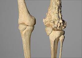
sessile (broad base) or pedunculated (narrow stalk) lesions found on the surface of bones
-
higher risk of malignant degeneration in sessile lesions
-
pedunculated lesions point away from the joint
-
-
continuity with native tissue
-
cortex of the lesion continuous with cortex of the native bone
-
medullary cavity of lesion continuous with medullary cavity of native bone
-
-
cartilage cap is usually radiolucent and involutes at skeletal maturity
-
nodules of metaplastic cartilage can occur within the bursa over cartilage caps
-
-
CT or MRI
-
used to better characterize lesions
-
HISTOLOGY
-
Characteristic histology
-
is similar to a normal physis with
-
cartilage cap consists of hyaline cartilage
-
well defined perichondrium around the cartilage cap
-
normal primary trabeculae
-
linear clusters of active chondrocytes
-
-
may have thin cartilage cap covers lesion
-
only 2-3 mm thick
-
thick cartilage caps imply growth but are not a reliable indicator of malignant degeneration in children
-
if cartilage cap becomes thicker as an adult, need to be concerned for chondrosarcoma transformation
-
-
DIFFERENTIALS
-
-
-
Differential of Osteochondroma
-
Surface lesions
-
May have similar chondrogenic histology
-
Treatment is Observation
-
Osteochondroma / MHE
-
o
-
o
-
o
-
Periosteal chondroma
-
o
-
o
-
Parosteal osteosarcoma
-
o
-
Periosteal osteosarcoma
-
o
-
Olliers / Maffucci
-
o
-
Chondrosarcoma
-
o
-
Paget's Disease
-
o
-
Enchondroma
-
o
-
Fibrous dysplasia
-
o
-
NOF
-
o
-
Eosinophillic granuloma
-
o
-
-
TREATMENT
-
Osteochondromas
-
nonoperative
-
observation alone
-
indications
-
asymptomatic or minimally symptomatic cases
-
-
-
-
operative
-
marginal resection at base of stalk, including cartilage cap
-
indications
-
symptomatic lesions
-
lesion may cause inflammation to surrounding tissue
-
lesion may be cosmetically displeasing
-
-
-
try to delay surgery until skeletal maturity
-
-
-
-
Multiple hereditary exostosis (MHE)
-
nonoperative
-
observation
-
indications
-
most patients do not require intervention prior to reaching skeletal maturity
-
-
-
-
operative
-
surgical excision of the osteochondroma
-
indications
-
dislocated radial heads
-
loss of forearm rotation
-
-
outcomes
- simple excision of the osteochondroma optimizes chance of improved motion
- simple excision of the osteochondroma optimizes chance of improved motion
-
-
-
-
Secondary chondrosarcoma
-
operative
- wide surgical resection
-
treat same as typical chondrosarcoma
-
- wide surgical resection
-
COMPLICATIONS
-
Pseudoaneurysm of the popliteal artery in the popliteal fossa
-
other vascular complications
-
vascular compression
-
true aneurysm
-
arterial thrombosis
-
venous thrombosis
-
-
-
Nerve compression
-
sciatic nerve
-
common peroneal nerve
-
atrophy of anterior and lateral compartment muscles of the leg
-
-
radial nerve
-
-
Tendon compression
-
lesions around the shoulder can give rise to
-
rotator cuff impingement
-
subscapularis tear
-
bicipital tendinitis
-
-
-
Chondrosarcoma
-
in adults, cartilage cap >2cm is associated with increased chance of malignancy
-
mean age of diagnosis, 31yrs
-
seldom in 1st decade or after 5th decade of life
-
-
-
Bursa formation
-
Recurrence
-
2-5% of cases after resection
-
Short-term X-ray surveillance is adequate unless symptomatic later
-
PROGNOSIS
-
Risk of malignant transformation is
-
<1% with solitary osteochondroma
-
~5-10% with MHE develop secondary chondrosarcoma
-