


-
SUMMARY
-
Scapula Fractures are uncommon fractures to the shoulder girdle caused by high energy trauma and associated with pulmonary injury, head injury, and increased injury severity scores.
-
Diagnosis can be made with plain radiographs and CT studies are helpful for fracture characterization and surgical planning.
-
Treatment is usually nonoperative with a sling. Surgical management is indicated for intra-articular fractures, displaced scapular body/neck fractures, open fractures, and those associated with glenohumeral instability.
-
-
EPIDEMIOLOGY
-
Incidence
-
rare
-
<1% of all fractures
-
3-5% of shoulder girdle fractures
-
-
-
Demographics
-
age
-
commonly between 25-50
-
-
males > females
-
-
Location
-
scapular body/spine = 45-50%
-
glenoid = 35%
-
glenoid neck = 25%
-
glenoid fossa/rim = 10%
-
often associated with impaction of humeral head into glenoid
-
-
acromion = 8%
-
coracoid = 7%
-
-
-
ETIOLOGY
-
Pathophysiology
-
mechanism of injury
-
high-energy trauma (80-90%)
-
motor vehicle collisions
-
account for >70% of scapula fractures
-
-
-
indirect trauma through fall on outstretched hand
-
glenohumeral dislocation
-
anterior dislocation leads to anterior rim fracture
-
posterior dislocation leads to posterior rim fracture
-
seizure
-
electric shock
-
-
-
-
-
Associated injuries (in 80-95%)
-
medical
-
thoracic injury (80%)
-
hemothorax/pneumothorax (>30%)
-
pulmonary contusion (>40%)
-
-
head injury (35-50%)
-
-
orthopaedic
-
rib fractures (53%)
-
ipsilateral extremity injury (50%)
-
ipsilateral clavicle fractures (25%)
-
-
spine fracture (26-30%)
-
pelvic ring/acetabular fractures (15%)
-
scapula fracture is important predictor
-
-
upper extremity vascular injury (11%)
-
subclavian and axillary arteries at risk
-
higher risk with scapulothoracic dissociation
-
-
brachial plexus injury (5-13%)
-
75% of brachial plexus injuries resolve
-
complete brachial plexus injuries less likely to resolve
-
-
-
-
-
-
ANATOMY
-
Osteology
-
scapular body
-
origin or insertion of 18 muscles
-
function to connect scapula to thorax, spine and upper extremity
-
-
large triangle shape with 4 major processes
-
scapular spine
-
osseous bridge separating supraspinatus and infraspinatus
-
spinoglenoid notch represents possible site of compression for suprascapular nerve
-
-
glenoid
-
represents articulating process on lateral scapula serving as socket for glenohumeral joint
-
pear-shaped and wider inferiorly from anterior to posterior
-
average 1-5º of retroversion and 15º superior tilt from scapular plane
-
fibrocartilaginous labrum deepens glenoid fossa by 50% to increase stability
-
-
acromion
-
articulates with clavicle to form acromioclavicular joint
-
formed by 3 ossification centers
-
pre acromion - tip
-
meso acromion - mid
-
meta acromion - base
-
-
-
coracoid process
-
has two secondary ossification centers that are open until around age 25 and should not be interpreted as fracture
-
angle of coracoid
-
tip of coracoid
-
-
muscular attachments
-
conjoint tendon
-
coracobrachialis
-
short head biceps
-
-
pectoralis minor
-
-
ligament attachments
-
coracoclavicular (CC) ligaments
-
most anterior CC ligament attachment is 25mm from tip of coracoid
-
-
coracoacromial ligament
-
-
-
-
-
-
Arthrology
-
glenohumeral joint
-
glenoid & labrum support humeral head to produce high degree of motion
-
stability provided by static and dynamic stabilizers
-
-
scapulothoracic joint
-
not a true joint but does represent an articulation between scapula and thorax
-
involved primarily in elevation and depression of shoulder as well as rotation and pro-/retraction
-
-
acromioclavicular (AC) joint
-
articulation of acromion and distal clavicle
-
supported by acromioclavicular ligaments (horizontal stability) and coracoclavicular ligaments (vertical stability)
-
-
8º of rotation occurs through acromioclavicular joint
-
-
superior shoulder suspensory complex
-
bone & soft tissue ring which provides connection of glenoid/scapula to axial skeleton
-
composed of 4 bony landmarks
-
distal clavicle
-
acromion
-
coracoid
-
glenoid
-
-
also composed of ligamentous complexes of acromioclavicular and coracoclavicular joints
-
-
-
Blood supply
-
contributions from anterior and posterior circumflex, scapular circumflex and suprascapular arteries
-
watershed area present in anterosuperior glenoid
-
-
Nervous system
-
scapula is intimately associated with brachial plexus
-
axillary nerve is at risk inferior to the glenoid as it runs from anterior to posterior
-
compression of suprascapular nerve at scapular notch leads to supraspinatus/infraspinatus weakness, with compression at the spinoglenoid notch leading only to infraspinatus weakness
-
-
Biomechanics
-
scapula contributes to glenohumeral rotation and abduction
-
1/3 of shoulder motion is scapulothoracic, 2/3 is glenohumeral
-
-
-
-
CLASSIFICATION
-
Classification is based on the location of the fracture and includes
-
scapular body fractures
-
usually described based on anatomic location
-
-
scapular neck fractures
-
look for associated AC joint separation or clavicle fracture
-
if occuring together, known as "floating shoulder"
-
-
-
glenoid fractures
-
Ideberg classification with Goss modification (below)
-
low inter- and intra-observer reliability and questionable association with management
-
-
AO-OTA classification
-
more reliable in diagnosis than Ideberg classification
-
-
-
acromial fractures
-
Kuhn classification
-
-
coracoid fractures
-
Ogawa classification - based on fracture proximity to CC ligaments
-
Eyres classification
-
-
scapulothoracic dissociation
-
-
-
Ogawa Coracoid Fracture Classification
-
Type I
-
Fracture occurs proximal to the coracoclavicular ligament
-
Type II
-
Fracture occurs towards the tip of the coracoid
-
-
-
Kuhn Acromial Fracture Classification
-
Type I
-
Nondisplaced or minimally displaced
-
Type II
-
Displaced but does not compromise the subacromial space
-
Type III
-
Displaced and compromises the subacromial space
-
-
-
Ideberg Classification of Glenoid Fracture
-
Type Ia
-
Anterior rim fracture
-
Type Ib
-
Posterior rim fracture
-
Type II
-
Fracture line through glenoid fossa exiting scapula inferiorly
-
Type III
-
Fracture line through glenoid fossa exiting scapula superiorly
-
Type IV
-
Fracture line through glenoid fossa exiting scapula medially through body
-
Type Va
-
Combination of types II and IV
-
Type Vb
-
Combination of types III and IV
-
Type Vc
-
Combination of types II, III, and IV
-
Type VI
-
Severe comminution
-
-
-
AO Classification for Glenoid Fractures
-
Fracture type
-
Subtype
-
Qualification
-
14F0: Extra-articular
-
Glenoid neck
-
14F1: Simple, intra-articular
-
1.1: anterior glenoid rim
-
1.2: posterior glenoid rim
-
1.3: transverse/short oblique
-
f: infraequitorial, single quadrant
-
r: supraequatorial, 2 quadrants
-
t: infraequitorial, 2 quadrants
-
i: infraequitorial
-
e: equitorial
-
p: supraequitorial
-
14F2: Multifragmentary
-
2.1: >= 3 articular fragments
-
2.2: central fracture-dislocation
-
14B: Extension into body
-
1: exits body at <=2 points
-
2: exits body at >=3 points
-
-
-
PRESENTATION
-
History
-
traumatic direct blow to shoulder or fall on outstretched arm
-
scapula fracture may be missed or diagnosed late in presence of other distracting, traumatic injuries
-
-
Symptoms
-
diffuse, severe shoulder pain
-
systemic symptoms
-
shortness of breath
-
chest wall pain
-
-
-
Physical exam
-
inspection
-
tenderness to palpation
-
shoulder diffusely
-
inaccurate in determining specific location of fracture
-
-
clavicle
-
spine
-
rib cage
-
-
evaluate for abnormal shoulder contour compared to contralateral site
-
look for open wounds or abrasions
-
soft tissue swelling may be significant
-
-
motion
-
acute active range of motion testing not recommended
-
likely to cause unnecessary pain
-
-
gentle passive range of motion can be useful in noting any blocks to motion
-
-
neurovascular
-
check motor and sensory function of nerves at risk
-
axillary
-
radial
-
median
-
ulnar
-
-
confirm symmetry of extremity pulses to contralateral side
-
-
-
-
IMAGING
-
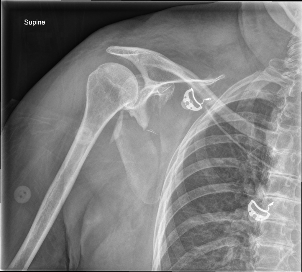
Radiographs
-
recommended views
-
true AP, grashey AP, scapular Y and axillary lateral view
-
AP chest radiograph
-
evaluate for pneumothorax
-
evaluate for widening of space between medial scapular border and spine
-
>1 cm indicates possible scapulothoracic dissociation
-
-
-
-
measurements
-
intra-articular step-off
-
lateral border offset (medialization)
-
glenopolar angle (measured on grashey AP)
-
angle connecting superior/inferior scapula and lateral border of scapula
-
normal considered 30-45º
-
-
scapular angulation
-
best seen on scapular Y radiograph
-
-
-
-
CT
-
indications
-
intra-articular fracture
-
significant displacement >1cm
-
may also help detect other thoracic/spine injuries
-
-
views
-
three-dimensional reconstruction better demonstrates fracture patterns
-
coronal and axial views useful to evaluate displacement, intra-articular step-off and medialization of glenoid
-
sagittal view useful to evaluate anterior-posterior displacement and angulation
-
-
-
MRI
-
indications
-
not regularly obtained but may be useful in some cases to evaluate the superior shoulder suspensory complex for ligamentous injury
-
-
-
-
DIFFERENTIAL
-
Os Acromiale
-
unfused secondary ossification centers (meso- and meta-acromion)
-
associated with impingement and rotator cuff symptoms and may be detected incidentally with trauma
-
-
-
-
TREATMENT
-
Nonoperative
-
sling for 2-3 weeks, followed by early motion
-
scapular body fractures
-
indications
-
indicated for vast majority of scapula fractures
-
90% are minimally displaced and acceptably aligned
-
-
outcomes
-
progressive deformity/displacement is possible during first 3 weeks
-
recommend serial weekly radiographs during this time
-
those associated with multiple underlying rib fractures or superior shoulder suspensory complex disruptions are more likely to displace
-
-
union at 6-8 weeks in most cases
-
most recover near-normal function
-
attributed to shoulder's capability for compensatory motion
-
-
poorer outcomes noted in patients with glenopolar angle <20º
-
-
-
scapular neck fractures
-
indications
-
translation <1 cm
-
angulation <40º
-
glenopolar angle >20º
-
no additional injury to superior shoulder suspensory complex
-
-
outcomes
-
true outcomes not well established
-
some reports of unsatisfactory results in ~30% of cases treated nonoperatively, while others note equivalent outcomes to surgical fixation
-
-
-
-
intra-articular glenoid fractures
-
indications
-
<4 mm step-off and less than 25% glenoid involvement
-
-
outcomes
-
with small fractures and minimal intra-articular step-off, nonoperative management results in excellent functional outcomes
-
risk of instability exists in rim fractures with larger degree of articular surface involvement
-
-
-
acromion fractures
-
indications
-
displacement <1 cm and no additional injury to superior shoulder suspensory complex
-
-
outcomes
-
good outcomes with Kuhn type I and II fractures which do not compromise subacromial space
-
-
-
coracoid fractures
-
indications
-
displacement <1 cm and no additional injury to superior shoulder suspensory complex
-
coracoid tip fractures distal to insertion of coracoclavicular (CC) ligaments, even if displacement is >1 cm (Ogawa II)
-
-
outcomes
-
good results and motion with both type I and II fractures meeting indications
-
-
-
-
-
Operative
-
open reduction internal fixation
-
indications (most are relative)
-
open fracture
-
scapular body fractures
-
medialization of lateral border > 20 mm
-
glenopolar angle < 20-22º
-
angulation > 40º
-
combination of medialization >15 mm and angulation >35º
-
-
scapular neck fracture
-
angulation > 40º
-
translation > 1 cm
-
glenopolar angle < 20-22º
-
"double disruption" of the superior shoulder suspensory complex (floating shoulder)
-
indicates unstable nature of bony/ligamentous ring
-
-
-
intra-articular glenoid fracture
-
> 20-25% anterior or posterior glenoid involvement with subluxation of humerus
-
can cause persistent glenohumeral instability
-
-
articular step-off > 4 mm
-
-
acromion fracture
-
displacement > 1cm
-
painful nonunion
-
subacromial impingement
-
double disruption of superior shoulder suspensory complex
-
-
coracoid fracture
-
displacement > 1 cm
-
painful nonunion
-
ipsilateral scapula fracture requiring fixation
-
Ogawa type I coracoid fracture extending into scapular body
-
double disruption of superior shoulder suspensory complex
-
-
-
techniques
-
screw(s)
-
percutaneous vs. open
-
-
plate(s) + screws(s)
-
arthroscopic-assisted
-
suture anchor repair vs. percutaneous screw fixation
-
useful in anterior/posterior glenoid rim fractures
-
-
-
-
outcomes
-
scapular body fractures
-
most return to having near-normal strength and symmetric range of motion
-
-
scapular neck fractures
-
good shoulder function and high union rates
-
complication rates up to 15%
-
-
intra-articular glenoid fractures
-
good to excellent subjective outcomes (pain, strength, and motion) in 80-95% of patients
-
higher rate of poor outcomes with concomitant chest and neurologic trauma
-
-
coracoid/acromion fractures
-
good outcomes in >85% of cases
-
high rates of union and full range of motion
-
-
some risk exists for requiring hardware removal
-
-
-
-
-
-
TECHNIQUES
-
Nonoperative (immobilization)
-
noninvasive but can lead to stiffness
-
technique
-
sling immobilization for 2-3 weeks
-
-
-
Open Reduction Internal Fixation (ORIF)
-
scapular body/neck fractures
-
approaches
-
straight posterior overlying glenohumeral joint
-
indicated in isolated displaced fractures
-
scapular neck
-
lateral scapular border
-
-
less extensile than Judet approach
-
-
Judet approach
-
indicated if multiple scapular borders need to be accessed
-
incision courses along spine of scapula and angles down vertebral scapula border in "L" shape
-
utilizes internervous plane between infraspinatus (suprascapular nerve) and teres minor (axillary nerve)
-
-
-
technique
-
can use 2.7 mm or 3.5 mm plates
-
locking plate technology may be advantageous given thin scapular bone, especially along vertebral border
-
reconstruction plates can be contoured around scapular spine and superomedial angle of scapula
-
-
complications
-
neurovascular injury
-
malunion
-
hardware failure
-
-
-
intra-articular glenoid fractures
-
approaches
-
deltopectoral approach
-
utilizes intermuscular plane between deltoid (axillary n.) and pectoralis major (medial/lateral pectoral n.)
-
indicated in fractures involving anterior glenoid with inferior extension (Ideberg II)
-
in cases of medial/inferior fracture extension into scapular body, posterior approach may be necessary
-
-
can be extended proximally to clavicle in cases where superior glenoid fracture extends to coracoid
-
-
posterior approach (detailed above)
-
displaced posterior glenoid rim fractures with intra-articular involvement
-
intra-articular glenoid fractures with inferior or medial extension into body not accessible anteriorly
-
-
lateral midaxial approach
-
incision just caudal to axilla in order to access inferior glenoid fractures
-
easier ability to instrument along inferior scapular neck
-
-
-
-
techniques
-
percutaneous fixation
-
if hardware is inserted percutaneously, arthroscopic assistance may be beneficial to ensure articular reduction
-
suture anchors can be used to advance labrum in cases of small bony defects
-
screw fixation can be used to fixate larger bony rim fragments
-
minifragment fixation recommended in most cases
-
-
-
-
open fixation
-
inferior glenoid fractures may be fixed with plate/screw(s) in buttress fashion
-
-
-
complications
-
post-traumatic arthritis
-
subscapularis failure
-
if anterior approach requires subscapularis take-down
-
-
recurrent glenohumeral instability
-
-
-
acromion fractures
-
approach
-
vertically based posterior incision centered over the scapular spine and posterior acromion
-
dissection taken down to deltoid and trapezius muscles and reflected off the scapular spine and posterior acromion
-
-
technique
-
proximal acromial fracture
-
2.7 or 3.5 mm lag screws placed perpendicular to fracture site if possible
-
2.4 or 2.7 mm reconstruction plate placed to neutralize fracture
-
-
distal acromial fracture
-
bone is very thin in this area
-
plate fixation may be difficult to obtain, although 2.0mm mini-fragment plate can function well
-
-
tension band technique can be considered
-
-
-
complications
-
hardware irritation/failure
-
-
-
coracoid fractures
-
approach
-
deltopectoral approach (detailed above)
-
retractor placed at base of coracoid to visualize fracture
-
-
-
technique
-
can carefully remove portion of the coracoacromial ligament and pectoralis minor attachment to better visualize the fracture bed
-
provisionally pin the coracoid with 1-2 Kirschner wires
-
fixation achieved with 1-2 bicortical 2.7 or 3.5 mm screws +/- washers
-
may also place quarter tubular buttress plate if needed
-
increased risk of requiring hardware removal
-
-
rarely, in Ogawa type II fractures requiring intervention, suture anchor can be placed in fracture bed and tip can be captured using a suture lasso technique
-
-
complications
-
neurovascular injury
-
hardware irritation
-
-
-
-
-
COMPLICATIONS
-
Post-traumatic glenohumeral arthritis
-
risk factors
-
intra-articular glenoid fracture with residual step-off/displacement
-
-
treatment
-
conservative management
-
NSAIDs, therapy, injections
-
-
shoulder arthroplasty (total vs. reverse)
-
-
-
Malunion
-
risk factors
-
higher degree of angulation, translation or medialization
-
more likely with nonoperative management
-
questionable effect on shoulder function
-
-
-
treatment
-
typically nonoperative depending on location of fracture and degree of deformity
-
If deformity involves glenoid, may be correctable with reverse total shoulder arthroplasty
-
-
-
Recurrent glenohumeral instability
-
risk factors
-
younger patients
-
larger degree of bone loss (anterior or posterior)
-
-
treatment
-
bony fixation (open or percutaneous)
-
arthroscopic vs. open suture anchor repair with labral advancement
-
more useful for smaller bony fragments which are not able to be fixated otherwise
-
-
-
-
Neurovascular injury
-
risk factors
-
scapulothoracic dissociation
-
iatrogenic injury during surgical dissection
-
deltopectoral approach
-
musculocutaneous n.
-
axillary n.
-
-
posterior/judet approach
-
axillary n.
-
suprascapular n.
-
circumflex scapular v.
-
posterior humeral circumflex v.
-
-
-
-
treatment
-
nerve injury after scapulothoracic dissociation
-
EMG 3-6 weeks after injury to assess extent of injury and degree of recovery
-
-
iatrogenic neurovascular injury
-
direct repair if possible
-
-
-
-