 اورژانس ارتوپدی و شکستگی ها


SUMMARY
-
Femoral Anteversion is a common congenital condition caused by intrauterine positioning which lead to increased anteversion of the femoral neck relative to the femur with compensatory internal rotation of the femur.
-
Diagnosis is made clinically with the presence of intoeing combined with an increase in internal rotation of the hip of greater than 70° with an accompanying decrease in external rotation of the hip of less than 20°.
-
Treatment is observation with parental reassurance as most cases resolve by age 10. Rarely, surgical management is indicated in the presence of less than 10° of hip external rotation in children greater than 10 years of age.
EPIDEMIOLOGY
-
Demographics
-
seen in early childhood (3-6 years)
-
twice as frequent in girls than boys
-
can be hereditary
-
-
Anatomic location
-
often bilateral
-
be cautious of asymmetric abnormalities
-
-
ETIOLOGY
-
Femoral anteversion is characterized by
-
increased anteversion of the femoral neck relative to the femur
-
compensatory internal rotation of the femur
-
lower extremity intoeing
-
-
There are three main causes of intoeing including
-
femoral anteversion (this topic)
-
metatarsus adductus (infants)
-
internal tibial torsion (toddlers)
-
-
Pathophysiology
-
a packaging disorders caused by intra-uterine positioning
-
most spontaneously resolve by age 10
-
-
Associated conditions
-
can be seen in association with other packaging disorders
-
DDH
-
metatarsus adductus
-
congenital muscular torticollis
-
-
ANATOMY
-
Is based on degree of anteversion of femoral neck in relation to the femoral condyles
-
at birth, normal femoral anteversion is 30-40°
-
typically decreases to normal adult range of 15° by skeletal maturity
-
minimal changes in femoral anteversion occur after age 8
-
PRESENTATION
-
Symptoms
-
parents complain of an intoeing gait in early childhood
-
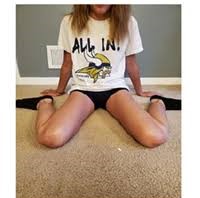
child classically sits in the W position (see above image)
-
knee pain when associated with tibial torsion
-
awkward running style
-
when extreme in an older child occasional functional limitations in sports and activities of daily living can occur
-
difficulty with tripping during walking or running activities
-
-
can be more symptomatic in those with neuromuscular diseases and brace-dependent walkers
-
secondary to lever-arm dysfunction and decreased compensatory mechanisms
-
-
-
Physical exam
- evaluation for intoeing
-
femoral anteversion
-
hip motion (tested in the prone position)
-
increased internal rotation of >70° (normal is 20-60°)
-
decreased external rotation of < 20° (normal 30-60°)
-
-
anteversion estimated on degree of hip IR when greater trochanter is most prominent laterally
-
trochanteric prominence angle test
-
-
patella internally rotated on gait evaluation
-
-
tibial torsion
-
look at thigh-foot angle in prone position
-
normal value in infants- mean 5° internal (range, −30° to +20°)
-
normal value at age 8 years- mean 10° external (range, −5° to +30°)
-
-
metatarsus adductus
-
adducted forefoot deformity, lateral border should be straight
-
a medial soft-tissue crease indicates a more rigid deformity
-
evaluate for hindfoot and subtalar motion
-
-
- evaluation for intoeing
IMAGING
-
Radiographs
-
recommended views
-
none required typically
-
-
-
CT or MRI
-
may be useful in measuring actual anteversion
-
TREATMENT
-
Nonoperative
- observation and parental reassurance
-
indications
-
most cases usually resolve spontaneously by age 10
-
-
technique
-
bracing, inserts, PT, sitting restrictions do not change natural history
-
-
- observation and parental reassurance
-
Operative
-
derotational femoral osteotomy
-
indications
-
< 10° of external rotation on exam in an older child (>10 yrs)
-
rarely needed
-
-
technique
-
typically performed at the intertrochanteric level
-
amount correction needed can be calculated by (IR-ER)/2
-
-
-
PROGNOSIS
-
Multiple studies have been unable to reveal any association with degenerative changes in the hip and knee when increased anteversion persists into adulthood