 Orthopedic emergency

Summary
-
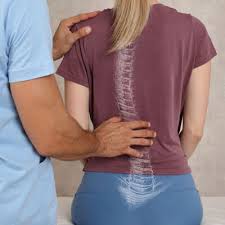
Adolescent Idiopathic Scoliosis is a coronal plane spinal deformity which most commonly presents in adolescent girls from ages 10 to 18.
-
Diagnosis is made with full-length standing PA and lateral spine radiographs.
-
Treatment can be observation, bracing, or surgical management depending on the skeletal maturity of the patient, magnitude of deformity, and curve progression.
Epidemiology
-
Incidence
-
most common type of scoliosis
-
incidence of 3% for curves between 10 to 20°
-
incidence of 0.3% for curves > 30°
-
-
-
Demographics
-
most commonly presents in children 10 to 18 yrs
-
10:1 female to male ratio for curves > 30°
-
1:1 male to female ratio for small curves
- right thoracic curve most common
-
left thoracic curves are rare and indicate an MRI to rule out cyst or syrinx
-
-
-
Etiology
-
Pathophysiology
-
unknown
-
potential causes
-
multifactorial
-
hormonal (melatonin)
-
brain stem
-
proprioception disorder
-
platelet
-
calmodulin
-
abnormal development of neurocentral synchodrosis (NCS)
- cartilaginous plate that forms between the centrum and posterior neural arches
-
closure occurs in characteristic order
-
cervical NCS by 5-6 years old
-
lumbar NCS by 11-12 years old
-
thoracic NCS by 14-17 years old
-
- cartilaginous plate that forms between the centrum and posterior neural arches
-
-
most have a positive family history
-
-
Curve Progression
-
risk factors for progression (at presentation)
- curve magnitude
-
before skeletal maturity
-
> 25° before skeletal maturity will continue to progress
-
-
after skeletal maturity
-
> 50° thoracic curve will progress 1-2° / year
-
> 40° lumbar curve will progress 1-2° / year
-
-
-
remaining skeletal growth
-
younger age
-
< 12 years at presentation
-
-
Tanner stage (< 3 for females)
- Risser Stage (0-1)
-
Risser 0 covers the first 2/3rd of the pubertal growth spurt
-
correlates with the greatest velocity of skeletal linear growth
-
- open triradiate cartilage
- peak growth velocity
-
is the best predictor of curve progression
-
in females it occurs just before menarche and before Risser 1 (girls usually reach skeletal maturity 1.5 yrs after menarche)
- most closely correlates with the Tanner-Whitehouse III RUS method of skeletal maturity determination
-
-
if curve is >30° before peak height velocity there is a strong likelihood of the need for surgery
-
-
-
curve type
-
thoracic more likely to progress than lumber
-
double curves more likely to progress than single curves
-
- curve magnitude
-
Classification
-
King-Moe Classification
-
five part classification to describe thoracic curve patterns and help guide surgeons implanting Harrington instrumentation
-
link to King-Moe classification (not testable)
-
-
Lenke Classification
-
more comprehensive classification based on PA, lateral, and supine bending films
-
helps to decide upon which curves need to be included within the fusion construct
-
link to Lenke classification (not testable)
-
Presentation
-
School screening
-
patients often referred from school screening where a 7° curve on scoliometer during Adams forward bending test is considered abnormal
-
7° correlates with 20° coronal plane curve
-
-
-
Physical exam
-
special tests
-
Adams forward bending test
-
axial plane deformity indicates structural curve
-
-
forward bending sitting test
-
can eliminate leg length inequality as cause of scoliosis
-
-
-
other important findings on physical exam
-
leg length inequality
-
midline skin defects (hairy patches, dimples, nevi)
-
signs of spinal dysraphism
-
-
shoulder height differences
-
truncal shift
-
rib rotational deformity (rib prominence)
-
waist asymmetry and pelvic tilt
-
cafe-au-lait spots (neurofibromatosis)
-
foot deformities (cavovarus)
-
can suggest neural axis abnormalities and warrant a MRI
-
-
asymmetric abdominal reflexes
-
perform MRI to rule out syringomyelia
-
-
-
Imaging
-
Radiographs
-
recommended views
-
standing PA and lateral
-
-
Cobb angle
-
> 10° defined as scoliosis
-
intra-interobserver error of 3-5°
-
-
spinal balance
-
coronal balance is determined by alignment of C7 plumb line to central sacral vertical line
-
sagittal balance is based on C7 plumb from center of C7 to the posterior-superior corner of S1
-
-
stable zone
-
between lines drawn vertically from lumbosacral facet joints
-
-
stable vertebrae
-
most proximal vertebrae that is most closely bisected by central sacral vertical line
-
-
neutral vertebrae
-
rotationally neutral (spinous process equal distance to pedicles on PA xray)
-
-
end vertebrae
-
end vertebra is defined as the vertebra that is most tilted from the horizontal apical vertebra
-
-
apical vertebrae
-
the apical vertebraeis the disk or vertebra deviated farthest from the center of the vertebral column
-
-
clavicle angle
-
best predictor of postoperative shoulder balance
-
-
-
MRI
-
should extend from posterior fossa to conus
-
purpose is to rule out intraspinal anomalies
-
indications to obtain MRI
- atypical curve pattern (left thoracic curve, short angular curve, apical kyphosis)
-
rapid progression
-
excessive kyphosis
-
structural abnormalities
-
neurologic symptoms or pain
-
foot deformities
-
asymmetric abdominal reflexes
-
a syrinx is associated with abnormal abdominal reflexes and a curve without significant rotation
- atypical curve pattern (left thoracic curve, short angular curve, apical kyphosis)
-
Treatment
- Based on skeletal maturity of patient, magnitude of deformity, and curve progression
-
Nonoperative
- observation alone
-
indications
-
cobb angle < 25°
-
-
technique
-
obtain serial radiographs to monitor for progression
-
-
- bracing
-
indication
-
cobb angle from 25° to 45°
- only effective for flexible deformity in skeletally immature patient (Risser 0, 1, 2)
-
goal is to stop progression, not to correct deformity
-
-
outcomes
- 50% reduction in need for surgery with compliant brace wear of at least 13 hours a day
-
poor prognosis with brace treatment associated with
-
poor in-brace correction
-
hypokyphosis (relative contraindication)
-
male
-
obese
-
noncompliant (effectiveness is dose-related)
-
- the number needed to treat (NNT) is four in highly compliant patients
-
Sanders staging system
-
predicts the risk of curve progression despite bracing to >50 degrees in Lenke type I and III curves
-
uses anteroposterior hand radiograph and curve magnitude to assess risk of progression despite bracing
-
- 50% reduction in need for surgery with compliant brace wear of at least 13 hours a day
-
- observation alone
-
Operative treatment
-
posterior spinal fusion
-
indications
- cobb angle > 45°
-
can be used for all types of idiopathic scoliosis
-
remains gold standard for thoracic and double major curves (most cases)
- cobb angle > 45°
-
-
anterior spinal fusion
-
indications
-
best for thoracolumbar and lumbar cases with a normal sagittal profile
-
-
-
anterior / posterior spinal fusion
-
indications
-
larges curves (> 75°) or stiff curves
-
young age (Risser grade 0, girls <10 yrs, boys < 13 yrs)
-
in order to prevent crankshaft phenomenon
-
-
-
-
Techniques
-
Bracing
- recommended for 16-23 hours/day until skeletal maturity or surgical intervention deemed necessary (actual wear minimum 12 hours required to slow progression)
-
brace types
-
curves with apex above T7
-
Milwaukee brace (cervicothoracolumbosacral orthosis)
-
extends to neck for apex above T7
-
-
-
apex at T7 or below
-
TLSO
-
Boston-style brace (under arm)
-
Charleston Bending brace is a curved night brace
-
-
-
bracing success is defined as <5° curve progression
-
bracing failure is defined
-
6° or more curve progression at orthotic discontinuation (skeletal maturity)
-
absolute progression to >45° either before or at skeletal maturity, or discontinuation in favor of surgery
-
-
skeletal maturity is defined as
-
Risser 4
-
<1cm change in height over 2 visits 6 months apart
-
2 years postmenarchal
-
- recommended for 16-23 hours/day until skeletal maturity or surgical intervention deemed necessary (actual wear minimum 12 hours required to slow progression)
-
Posterior spinal fusion
-
fusion levels
-
goals
-
fusion should include enough levels to adequately maintain sagittal and coronal balance while being as minimal as safely possible to preserve motion
-
typical fusion from proximal end vertebra to one or two levels cephalad to the stable vertebra
-
double and triple major curves fuse to the distal end vertebra
-
-
Harrington technique
-
recommends one level above and two levels below the end vertebrae if these levels fall wilthin the stable zone
-
-
Moe technique
-
recommends fusion to the neutral vertebrae
-
-
Lenke technique
-
recommends including all major curves in the fusion and minor curves that are not flexible or are kyphotic
-
-
L5 level
-
Cochran found increase incidence of low back pain with fusion to L5, and to a lesser extent L4.
-
therefore, whenever possible, avoid fusion to L4 and L5
-
-
-
pelvis
-
it is almost never required to fuse to the pelvis in idiopathic scoliosis
-
-
-
pedicle screw fixation
-
screw insertional torque correlates with resistance to screw pullout
-
resistance to screw pullout increases by
- undertapping by 1mm
- undertapping by 1mm
-
-
curve correction
-
segmental pedicle screw fixation allows increased coronal plane correction while lessening the need for anterior releases
-
-
-
ASF with instrumentation
-
advantage
-
better correction while saving lumbar fusion levels
-
-
disadvantage
- increased risk of pseudarthrosis when thoracic hyperkyphosis is present
- increased risk of pseudarthrosis when thoracic hyperkyphosis is present
-
fusion levels
-
typically fuse from end vertebra to end vertebra
-
-
-
Neurologic Monitoring
-
monitoring with somatosensory-evoked potentials (SSEPs) and/or motor-evoked potentials (MEPs) is now the standard of care
-
motor-evoked potentials can provide an intraoperative warning of impending spinal cord dysfunction
-
-
neurologic event defined as drop in amplitude of > 50%
-
if neurologic injury occurs intraoperatively consider
-
check for technical problems
-
check blood pressure and elevate if low
-
check hemoglobin and transfuse as necessary
-
lessen/reverse correction
-
administer Stagnaras wake up test
-
remove instrumentation if the spine is stable
-
-
Complications
-
Neurologic injury
-
paraplegia is 1:1000
-
increased risk with kyphosis, excessive correction, and sublaminar wires
-
- Pseudoarthrosis (1-2%)
-
presents as late pain, deformity progression, and hardware failure
-
an asymptomatic pseudarthrosis with no pain and no loss of correction should be observed
-
-
-
Infection (1-2%)
-
presents as late pain
-
incision often looks clean
-
Propionibacterium acnes most common organism for delayed infection (requires 2 weeks for culture incubation)
-
attempt I&D with maintenance of hardware if not loose and within 6 months
-
-
Flat back syndrome
-
early fatigability and back pain due to loss of lumbar lordosis
-
rare now that segmental instrumentation addresses sagittal plane deformities
-
decreased incidence with rod contouring in the sagittal plane and compression/distraction techniques
-
-
treat with revision surgery utilizing posterior closing wedge osteotomies
-
anterior releases prior to osteotomies aid in maintenance of correction
-
-
-
Crankshaft phenomenon
-
rotational deformity of the spine created by continued anterior spinal growth in the setting of a posterior spinal fusion
-
can occur in very young patients when PSF is performed alone and the anterior column is allowed continued growth
-
avoided by performing anterior diskectomy and fusion with posterior fusion in very young patients
-
-
-
SMA syndrome (superior mesenteric artery [SMA] syndrome)
-
compression of 3rd part of duodenum due to narrowing of the space between SMA and aorta
-
SMA arises from anterior aspect of aorta at level of L1 vertebrae
-
presents with symptoms of bowel obstruction in first postoperative week
-
associated with electrolyte abnormalities
-
nausea, bilious vomiting, weight loss
-
-
risk factors
-
height percentile <50%; weight percentile < 25%
-
sagittal kyphosis
-
-
treat with NG tube and IV fluids
-
-
Hardware failure
-
late rod breakage can signify a pseudarthrosis
-
-
Emergency department visits
-
most often for minor medical complaints
-
associated with older age at the time of surgery and more fusion levels
-
-
Prognosis
-
Natural history
- increased incidence of acute and chronic pain in adults if left untreated
-
curves > 90° are associated with cardiopulmonary dysfunction, early death, pain, and decreased self image
- increased incidence of acute and chronic pain in adults if left untreated