 Orthopedic emergency


SUMMARY
-
Equinovarus Foot is an acquired foot deformity commonly seen in pediatric patients with cerebral palsy, spina bifida, and Duchenne Muscular Dystrophy that present with a equinovarus foot deformity.
-
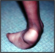
Diagnosis is made clinically with presence of an inverted heel with a supinated forefoot, often associated with pain and callous formation along the lateral border of the foot.
-
Treatment ranges from bracing to tendon transfers to osteotomies depending on the underlying etiology, severity of deformity, and rigidity of contracture.
EPIDEMIOLOGY
-
Incidence
-
common foot deformity seen with
-
cerebral palsy (usually spastic hemiplegia)
-
Duchenne muscular dystrophy
-
residual clubfoot deformity
-
spina bifida
-
tibial deficiency (hemimelia)
-
though this condition is very rare
-
-
-
ETIOLOGY
-
-
Pathophysiology
-
pathomechanics
-
imabalance of invertors and evertors (invertors overpower the evertors)
-
relative overpull of
-
tibialis posterior and/or
-
tibialis anterior
-
gastoc-soleus complex
-
-
example: in cerebral palsy
-
the causative muscles for the varus are the
-
anterior tibialis (AT) in 1/3 of patients
-
posterior tibialis (PT) in 1/3 and
-
both the AT and PT in the remaining 1/3
-
-
-
-
foot deformity muscle imbalance overview
-
-
PRESENTATION
-
Symptoms
-
pain
-
painful weight bearing over the lateral border of the foot
-
-
instability
-
during stance phase
-
results in shortened single limb stance
-
-
poor shoe and/or brace fitting and shoe wear problems
-
-
Physical Exam
-
inspection
-
inverted heel (tibialis posterior typically implicated)
-
supinated forefoot (tibialis anterior)
-
callous and pain along lateral border
-
intoeing gait (foot progression angle is more internal than knee progression angle)
-
-
provocative tests
-
active dorsiflexion of foot
-
if foot supinates with dorsiflexion, the anterior tibialis is implicated
-
-
confusion test
-
indications
-
used in those with poor selective motor control, as in CP, and cannot dorsiflex foot when asked)
-
-
method
-
patient performs active hip flexion (with or without resistance) while seated
-
results in ankle dorsiflexion due to mass action pattern of leg
-
if the foot supinates with dorsiflexion, the tibialis anterior is likely a contributing to the varus deformity
-
-
-
-
Coleman block test
-
indications
-
to test rigidity of the varus deformity
-
do not do this in children with limited balance such as CP
-
-
method
-
patient stands on a block with the first ray off the block
-
if the varus corrects, the deformity is flexible
-
-
-
manual manipulation of the hindfoot
-
can be used to asses rigidity of the varus deformity
-
passive eversion of the hindfoot past neutral demonstrates that the varus deformity is flexible
-
-
-
IMAGING
-
Radiographs
-
recommended views
-
AP + lateral of foot
-
-
findings
-
forefoot adduction is seen on the AP radiograph
-
the talus and calcaneus are more parallel than in typical feet
-
one can often "look down" the sinus tarsi through a visual hole there
-
the calcaneus looks foreshortened on the lateral view
-
the metatarsals are often "stacked" on the lateral view (instead of being in line with one another)
-
stress fractures along the fourth and/or fifth metatarsal bases can develop secondary to repetitive load along the lateral border of the foot.
-
-
STUDIES
-
Dynamic EMG
-
may be useful in distinguishing whether tibialis anterior and/or tibialis posterior is/are causing the varus in CP
-
TREATMENT
-
Nonoperative
-
ankle foot orthosis (AFO)
-
helps provide stability for the foot and a more stable base of support during gait
-
should have a "wrap around" hindfoot component of the brace to help control the varus and minimize pressure points
-
-
serial casting
-
indication
-
rigid deformity
-
-
-
botulinum toxin injection into tibialis posterior and/or gastrocnemius
-
indication
-
flexible or dynamic deformities
-
desire to delay surgery
-
-
-
-
Operative
-
gastrocnemius recession or tendoachilles lengtheing (TAL) for equinus
-
indications
-
fixed equinus unresponsive to non-operative measures
-
gastrocnemius recession should be performed if the anke can be brought to neutral or above neutral with the knee flexed and hindfoot inverted, but not when the knee is extended
-
TAL should be performed if the ankle can not be dorsiflexed to neutral with the knee flexed or extended
-
-
-
split-posterior tibialis tendon transfer [SPOTT] or posterior tibial tendon lengthening (PTTL)
-
indications
-
soft tissue balancing is required if varus is flexible or rigid
-
varus foot recalcitrant to non-operative measures and posterior tibialis contributing to varus (dynamic EMG, when available is helpful)
-
tibialis posterior spastic in both stance and swing phase (continous activity)
-
common patient: spastic hemiplegia in ages 5 to 7 years old
-
-
technique
-
SPOTT
-
reroute half of tendon laterally and insert into peroneus brevis
-
-
PTTL
-
fractional lengthening of the tendon in the distal third of the lower leg
-
-
either PTTL or SPOTT may be combined with SPLATT
-
-
outcomes
-
results for both surgeries are good, without clear indications for transfer versus lengthening
-
-
-
split-anterior tibialis tendon transfer [SPLATT]
-
indications
-
overactive anterior tibialis on EMG
-
when anterior tibialis contributes to varus foot, whether flexible or rigid varus deformity
-
-
technique
-
split anterior tibialis transfer to cuboid, peroneus tertius, or peroneus brevis
-
may be combined with SPOTT or PTTL
-
-
-
calcaneal osteotomy
-
indications
-
required for a rigid hindfoot varus deformity
-
-
technique
-
lateral closing wedge osteotomy (Dwyer) to incur valgus to the heel, OR
-
lateral calcaneal sliding osteotomy to correct the varus
-
typically combined with soft tissue balancing (as above)
-
-
-
COMPLICATION
-
Overcorrection (resultant valgus deformity)
-
increased risk in
-
children who undergo surgery at younger age
-
children with diplegia (as oppose to hemiplegia)
-
-
-
Wound complications
-
most common with calcaneal osteotomy lateral incision
-
risk decreased by using absorbable suture
-
-
Hardware Pressure sores/ulcers
-
from buttons on bottom of foot (from SPLATT to cuboid)
-
has led some surgeons to always transfer SPLATT to peroneus tertius or brevis
-