 Orthopedic emergency


-
SUMMARY
-
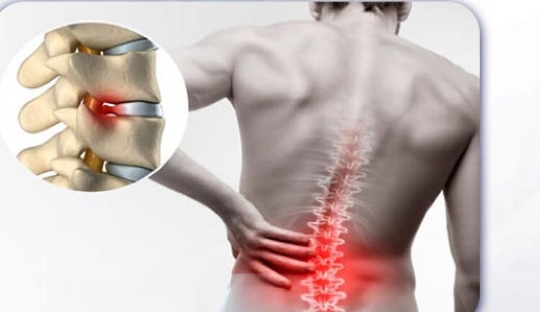
Lumbar Disc Herniation is a very common cause of low back pain and unilateral leg pain, known as radiculopathy. In rare cases a large disc herniation can lead to Cauda Equina Syndrome which requires emergent diagnosis and treatment.
-
Diagnosis is made clinically and confirmed with an MRI studies of the lumbar spine.
-
Treatment for radicular leg pain is initially nonoperative with oral medications and physical therapy. Surgical microdiscectomy is only indicated for severe pain and/or motor deficit that have failed to respond to nonoperative management. Treatment for Cauda Equina Syndrome in contrast is emergent microdiscectomy within 48 hours.
-
-
EPIDEMIOLOGY
-
Incidence
-
peak incidence is 4th and 5th decades
-
lifetime prevalence of 10%
-
only ~5% become symptomatic
-
-
Demographics
-
3:1 male:female ratio
-
-
Location
-
L5/S1 most common level
-
95% involve L4/5 or L5/S1 levels
-
-
-
ETIOLOGY
-
Pathoanatomy
-
recurrent torsional strain leads to tears of the outer annulus which leads to herniation of nucleus pulposis
-
lateral edge of posterior longitudinal ligament weakest region
-
common site for posterolateral/paracentral disc herniations
-
-
sinuvertebral nerves provide pain innervation to the posterior annulus
-
mediate vertebrogenic back pain that precedes or accompanies disc herniation
-
-
-
Pathophysiology
-
cellular senescence of fibrochondrocytes leads to loss of proteoglycan production leading to disc height loss
-
loss of height causes increased strain on the annulus fibrosus
-
increased strain leads to fissures of the annulus fibrils
-
-
annular tears compromise hoop stresses that act against the deforming forces of the nucleus pulposus
-
nucleus pulposus herniates through tear
-
younger, well-hydrated discs more likely to herniate
-
pediatric patients may have Salter-Harris II fracture of the ring apophysis
-
-
older, desiccated discs less likely to herniate
-
-
sciatica symptoms result from combined mechanical compression and associated inflammation
-
not all patients with mechanical compression develop symptoms
-
TNF-α, MMP, NO, PE2, and IL-6 are implicated in nerve irritation leading to radiculopathy
-
weak evidence to support DMARDs for treatment
-
-
-
-
-
-
ANATOMY
-
Complete intervertebral disc anatomy and biomechanics
-
Disc composition
-
annulus fibrosis
-
composed of type I collagen, water, and proteoglycans
-
15-25 sheets of lamellae
-
-
characterized by extensibility and tensile strength
-
high collagen / low proteoglycan ratio (low % dry weight of proteoglycans)
-
-
-
nucleus pulposus
-
composed of type II collagen, water, and proteoglycans
-
characterized by compressibility
-
low collagen / high proteoglycan ratio (high % dry weight of proteoglycans)
-
proteoglycans interact with water and resist compression
-
-
a hydrated gel due to high polysaccharide content and high water content (88%)
-
disc height dependent on the degree of hydration
-
-
-
avascular structure
-
nutrients supplied by diffusion from the end plates
-
-
-
-
Nerve root anatomy
-
key difference between cervical and lumbar spine is
-
pedicle/nerve root mismatch
-
cervical spine C6 nerve root travels under C5 pedicle (mismatch)
-
lumbar spine L5 nerve root travels under L5 pedicle (match)
-
extra C8 nerve root (no C8 pedicle) allows transition
-
-
horizontal (cervical) vs. vertical (lumbar) anatomy of nerve root
-
because of vertical anatomy of lumbar nerve root a paracentral and foraminal disc will affect different nerve roots
-
because of horizontal anatomy of cervical nerve root a central and foraminal disc will affect the same nerve root
-
-
-
-
-
CLASSIFICATION
-
Location Classification
-
central prolapse
-
often associated with back pain only
-
may present with cauda equina syndrome which is a surgical emergency
-
-
posterolateral (paracentral)
-
most common (90-95%)
-
PLL is weakest here
- affects the traversing/descending/lower nerve root
-
at L4/5 affects L5 nerve root
-
-
- foraminal (far lateral, extraforaminal)
-
less common (5-10%)
- affects exiting/upper nerve root
-
at L4/5 affects L4 nerve root
-
-
herniated disc material directly compresses dorsal root ganglion
- can manifest with more severe pain than traditional posterolateral disc herniation
- can manifest with more severe pain than traditional posterolateral disc herniation
-
-
axillary
-
can affect both exiting and descending nerve roots
-
-
-
Morphology classification
-
protrusion
-
eccentric bulging with an intact annulus
-
-
extrusion
-
disc material herniates through annulus but remains continuous with disc space
-
-
sequestered fragment (free)
-
disc material herniates through annulus and is no longer continuous with disc space
-
prone to proximal or distal migration
-
-
-
Containment classification
-
contained
-
disc material is contained beneath the posterior longitudinal ligament
-
-
uncontained
-
disc material passes dorsal to the posterior longitudinal ligament
-
-
-
Timing classification
-
acute
-
herniations present < 3-6 months
-
important consideration given surgical outcomes are associated with chronicity
-
-
-
chronic
-
herniations present >6 months
-
-
-
-
PRESENTATION
-
History
-
sudden onset of pain after lifting a heavy object
-
occupational exposure
-
prolonged sitting with lateral bending and rotation in the presence of vibrational energy
-
-
symptomatic improvement lying supine with knees and hips flexed
-
especially with lower lumbar disc herniations
-
-
-
Symptoms
-
can present with symptoms of
-
axial back pain (low back pain)
-
this may be discogenic or mechanical in nature
-
can precede herniation
-
-
radicular pain (buttock and leg pain)
-
often worse with sitting, improves with standing
-
symptoms worsened by coughing, valsalva, sneezing
-
pain not worsened with ambulation
-
-
cauda equina syndrome (present in 1-10%)
-
bilateral leg pain
-
LE weakness
-
saddle anesthesia
-
bowel/bladder symptoms
-
-
-
- Physical exam
-
inspection
-
limited lumbar range of motion
-
often the pain is the limiting factor
-
-
patient leaning away from side of radiculopathy
-
effort to increase the size of the neuroforamen
-
-
-
palpation
-
spasms of the paraspinal musculature
-
nonspecific
-
-
associated tenderness in the paraspinal musculature
-
nonspecific
-
-
-
motor exam & reflexes
-
see lower extremity neuro exam
-
L3 radiculopathy
- hip adduction weakness
-
knee extension weakness
-
dermatomal pain in the anteromedial thigh
- hip adduction weakness
-
L4 radiculopathy
-
ankle dorsiflexion weakness (L4 > L5)
-
decreased patellar reflex
-
dermatomal pain in the lateral thigh, crossing the knee, to medial foot
-
-
L5 radiculopathy
-
EHL weakness (L5)
-
manual testing
-
-
ankle dorsiflexion weakness (L4 > L5 contribution)
-
test by having patient walk on heels
-
- ankle inversion weakness
- hip abduction weakness (L5)
-
have patient lie on side on exam table and abduct leg against resistance
-
-
dermatomal pain in anterolateral leg and dorsum of foot
-
-
S1 radiculopathy
-
ankle plantar flexion weakness (S1)
-
have patient do 10 single leg toes stands
-
-
decreased Achilles tendon reflex
-
dermatomal pain in posterior calf and lateral foot
-
-
-
-
provocative tests
-
straight leg raise (Lasegue's sign)
-
a tension sign for L4, L5 and S1 nerve root
-
technique
-
can be done sitting or supine
-
reproduces pain and paresthesia in leg at 30-70 degrees hip flexion
-
-
sensitivity/specificity
-
most important and predictive physical finding for identifying who is a good candidate for surgery
-
-
-
contralateral SLR
-
crossed straight leg raise is less sensitive but more specific
-
-
femoral nerve stretch test (Wasserman sign)
-
tension sign for L2 and L3
-
performed in prone position
-
knee flexed and hip exteneded
-
reproduction of pain in anterior thigh is considered positive
-
-
-
Braggard's sign
-
perform SLR to the point of exacerbation
-
lower leg just to the point where pain recedes
-
ankle dorsiflexion causes exacerbated pain
-
-
-
Bowstring sign
-
SLR aggravated by compression on popliteal fossa
-
-
Kernig test
-
pain reproduced with neck flexion, hip flexion, and leg extension
-
-
Naffziger test
-
pain reproduced by coughing, which is instigated by lying patient supine and applying pressure on the neck veins
-
-
Milgram test
-
pain reproduced with straight leg elevation for 30 seconds in the supine position
-
-
-
gait analysis
-
Trendelenburg gait
-
due to gluteus medius weakness which is innervated by L5
-
-
-
-
-
IMAGING
-
Radiographs
-
recommended views
-
AP and lateral radiographs
-
helpful for surgical localization
-
identify anomalous vertebrae (sacralized L5 or lumbarized S1)
-
-
-
-
optional views
-
flexion-extension
-
identifies instability
-
if present can changes surgical plan to involve fusion
-
-
-
-
findings
-
most often normal
-
abnormal findings
-
loss of lordosis (spasm)
-
loss of disc height
-
especially at the involved level
-
-
lumbar spondylosis (degenerative changes)
-
facet hypertrophy
-
disc space collapse
-
peridiscal osteophytes
-
-
sciatic scoliosis
-
convex or concave list to the ipsilateral side of herniation
-
-
-
-
sensitivity
-
poor sensitivity for identifying disc herniation
-
more often used as a screening tool for other pathology prior to proceeding with MRI
-
-
-
CT myelogram
-
indications
-
patients unable to obtain MRI
-
pacemaker
-
-
-
views
-
sagittal and coronal reconstructions demonstrate compression of the thecal sac
-
-
findings
-
myelography filling defect at the level of herniation
-
a calcified disc may be visible
-
-
sensitivity
-
93% accurate at detecting associated surgical pathology
-
unable to detect foraminal or extraforaminal herniations
-
-
-
MRI without gadolinium
-
indications for obtaining an MRI
-
pain lasting > one month and not responding to nonoperative management or
-
red flags are present
-
infection (IV drug user, h/o of fever and chills)
-
tumor (h/o or cancer)
-
trauma (h/o car accident or fall)
-
cauda equina syndrome (bowel/bladder changes)
-
-
-
modality of choice for diagnosis of lumbar disc herniations
-
highly sensitive and specific
-
helpful for preoperative planning
-
useful to differentiate from synovial facet cysts
-
-
however high rate of abnormal findings on MRI in normal people
-
need to correlate MRI findings with symptoms and physical exam findings
-
-
views
-
sagittal and coronal T2 reconstructions
-
localize the level and side of the herniation
-
location anatomic location (central vs paracentral vs foraminal vs extraforaminal)
-
-
-
-
MRI with gadolinium
-
indications
-
useful for revision surgery
-
-
findings
-
allows to distinguish between post-surgical fibrosus (enhances with gadolinium) vs. recurrent herniated disc (does not enhance with gadolinium)
-
-
-
-
TREATMENT
-
Nonoperative
- rest and physical therapy, anti-inflammatory medications, and limited narcotics
-
indications
-
first line of treatment for most patients with disc herniation
-
new-onset radicular pain
-
no significant motor weakness
-
absence of cauda equina syndrome
-
no bowel/bladder incontinence
-
-
-
outcomes
-
90% improve without surgery
-
positive predictors of good outcomes with nonoperative treatment
-
higher level of education
-
-
-
- selective nerve root corticosteroid injections
-
indications
-
second line of treatment if therapy and medications fail
-
usually after 6 weeks
-
-
-
outcomes
-
leads to long lasting improvement in ~ 50% (compared to ~90% with surgery)
-
results best in patients with extruded discs as opposed to contained discs
-
no difference in pain relief using lidocaine with and without steroids
-
-
- rest and physical therapy, anti-inflammatory medications, and limited narcotics
-
Operative
-
laminotomy and discectomy (microdiscectomy)
- indications
-
persistent disabling pain lasting more than 6 weeks that have failed nonoperative options (and epidural injections)
-
timing of appropriate nonoperative treatment varies
-
better surgical outcomes if addressed within 2 months
-
- progressive and significant weakness
-
cauda equina syndrome
-
-
rehabilitation
- patients may return to medium to high-intensity activity at 4 to 6 weeks
- patients may return to medium to high-intensity activity at 4 to 6 weeks
-
outcomes
-
outcomes with surgery compared to nonoperative
- improvement in pain and function greater with surgery
-
early and sustained pain relief out to 2 years
-
equal likelihood of receiving disability at 5 years
- improvement in pain and function greater with surgery
-
positive predictors for good outcome with surgery
-
leg pain is chief complaint
-
positive straight leg raise
-
weakness that correlates with nerve root impingement seen on MRI
-
married status
- progressively worsening symptoms prior to surgery
- professional athletes
-
younger age, greater number of games played prior to injury
-
-
paracentral and foraminal herniations
-
central and extraforaminal associated with worse outcomes
-
-
herniation at caudal levels
-
L5-S1 results in better outcomes than L2-3
-
-
-
negative predictors for good outcome with surgery
- worker's compensation
-
WC patients have less relief from symptoms and less improvement in quality of life with surgical treatment
-
-
smokers
-
chronic headaches
-
depression
- worker's compensation
-
- indications
-
far lateral microdiskectomy
-
indications
-
for far-lateral disc herniations
-
-
-
-
-
TECHNIQUES
-
Rest and physical therapy, anti-inflammatory medications, and limited narcotics
-
bedrest followed by progressive activity as tolerated
-
historical treatment
-
most modern protocols involve immediate activity with modification to avoid pain exacerbation
-
-
-
medications
-
NSAIDS
-
muscle relaxants (more effective than placebo but have side effects)
-
oral steroid taper
-
modest but significant improvement in function, no significant improvement in pain
-
-
narcotic medications
-
typically avoided due to complication profile
-
dependence
-
difficult post-op pain control
-
worse outcomes following surgical treatment
-
-
if used, usually for a short period (2-3 days) in the acute setting
-
-
-
physical therapy
-
typically initiated three weeks after symptom onset
-
extension exercises are extremely beneficial
-
traction
-
chiropractic manipulation
-
should be performed with care
-
-
-
-
Selective nerve root corticosteroid injections
-
epidural
-
selective nerve block
-
can be therapeutic and diagnostic
-
useful in case of diagnostic dilemmas
-
-
-
-
Laminotomy and discectomy (microdiscectomy)
-
various techniques available
-
most techniques can be performed in a "minimally invasive" fashion
-
can be done with small incision or through "tube" access
-
open technique using a crank (McCulloh) retractor
-
-
discectomy performed through microscope or loupe magnification
-
no difference in outcomes between the two
-
-
endoscopic techniques available
-
provide smaller incisions
-
-
similar outcomes between all techniques surgical techniques
-
fragment excision vs extended disc space curettage (subtotal discectomy)
-
lower long term back pain with fragment excision
-
higher reherniation rates with fragment excision at 2-years follow-up
-
-
-
-
Far lateral microdiskectomy
-
utilizes a paraspinal approach of Wiltse
- can also be done with tubular or crank retractors
- can also be done with tubular or crank retractors
-
-
-
COMPLICATIONS
-
Dural tear
-
occurs in 0-4% of cases
-
treatment
-
if have tear at time of surgery then perform water-tight repair
-
has not been shown to adversely affect long term outcomes
-
-
-
- Recurrent HNP
-
defined as recurrent sciatica at the same operated level
-
pain-free interval of 6 months prior to recurrence of symptoms
-
pathology can be ipsilateral to contralateral to the index presentation
-
-
recurrence rate 5-15%
-
revision rate at 8-year follow-up is 15% according to the SPORT trial
-
risk factors protective against recurrent herniation
-
discrete herniations
-
small annular defects (<6 mm)
-
-
-
treatment
-
can treat nonoperatively initially
-
revision microdiscectomy in patients with persistent symptoms
- outcomes for revision discectomy have been shown to be as good as for primary discectomy
- outcomes for revision discectomy have been shown to be as good as for primary discectomy
-
-
-
Wound infections
-
occurs in up to 3% of cases
-
epidural abscess in 0.3% of cases
-
-
risk factors
-
microscope usage proposed as a source of infection
-
some date refutes this claim
-
-
-
treatment
-
superficial infections
-
treat with local wound care and antibiotics
-
-
deep infections
-
surgical I&D
-
-
-
-
Epidural fibrosis
-
scarring the compresses the dura leading to radicular symptoms
-
associated with poor outcomes following revision surgery
-
persistent back pain
-
patients 3.2 times more likely to suffer from recurrent radiculopathy
-
-
-
MRI may demonstrate retraction of the dura on the side of the lesion
-
-
Pyogenic discitis
-
occurs in 2.3% of cases
-
treatment
-
IV antibiotics +/- surgical I&D
-
-
-
Chronic low back pain
- not completely understood but central sensitization may be a factor
-
amplification of neural signaling within the central nervous system (CNS) that elicits pain hypersensitivity.
-
-
Modic changes on MRI imaging are associated with post-operative back pain
- Pain diagrams may be useful in identifying patients with an increased likelihood of pain sensitization, psychosocial load, and utilizing pain management resources
- not completely understood but central sensitization may be a factor
-
Vascular catastrophe
-
exceedingly rare
-
caused by breaking through anterior annulus and injuring vena cava/aorta
-
treatment
-
immediate recognition of complication followed by coordinated repair by vascular service
-
-
-
Instability
-
due to over resection of lamina and pars interarticularis
-
not all patients are symptomatic
-
treatment
-
instrumentation and fusion of the affected segment
-
-
-
-
PROGNOSIS
-
Natural history
-
90% of patients will have improvement of symptoms within 3 months without substantial medical treatment
-
patients less likely to improve if still symptomatic after 6 weeks
-
-
factors associated with good outcomes with nonoperative treatment
-
lack of radiculopathy
-
-
factors associated with worse outcomes with nonoperative treatment
-
obese patients (BMI >30)
-
symptoms present >6 months prior to starting treatment
-
-
- Size of herniation decreases over time (reabsorbed)
-
sequestered disc herniations show the greatest degree of spontaneous reabsorption
-
macrophage phagocytosis and enzymatic degradation is the mechanism of reabsorption
-
-
Factors associated with favorable surgical outcomes
-
severe preoperative leg pain
-
shorter symptom duration
-
younger age
-
increased preoperative physical activity
-
-
Surgical treatment is equivalent to nonsurgical treatment in the long term
-
surgery provides faster pain relief
-
-